
A A Rare Case Report on Lhermitte-Duclos Disease: An Intraoperative and Radiopathological Diagnosis
DOI:
https://doi.org/10.21276/apalm.3331Keywords:
Lhermitte-Duclos Disease, Cerebellum, HistopathologyAbstract
Background: Lhermitte-Duclos Disease (LDD), also known as dysplastic gangliocytoma of the cerebellum, is a very rare tumor of the cerebellum, with only about 300 cases reported worldwide. It is a slow-growing benign tumor causing diffuse enlargement of the cerebellum due to granular layer hypertrophy.
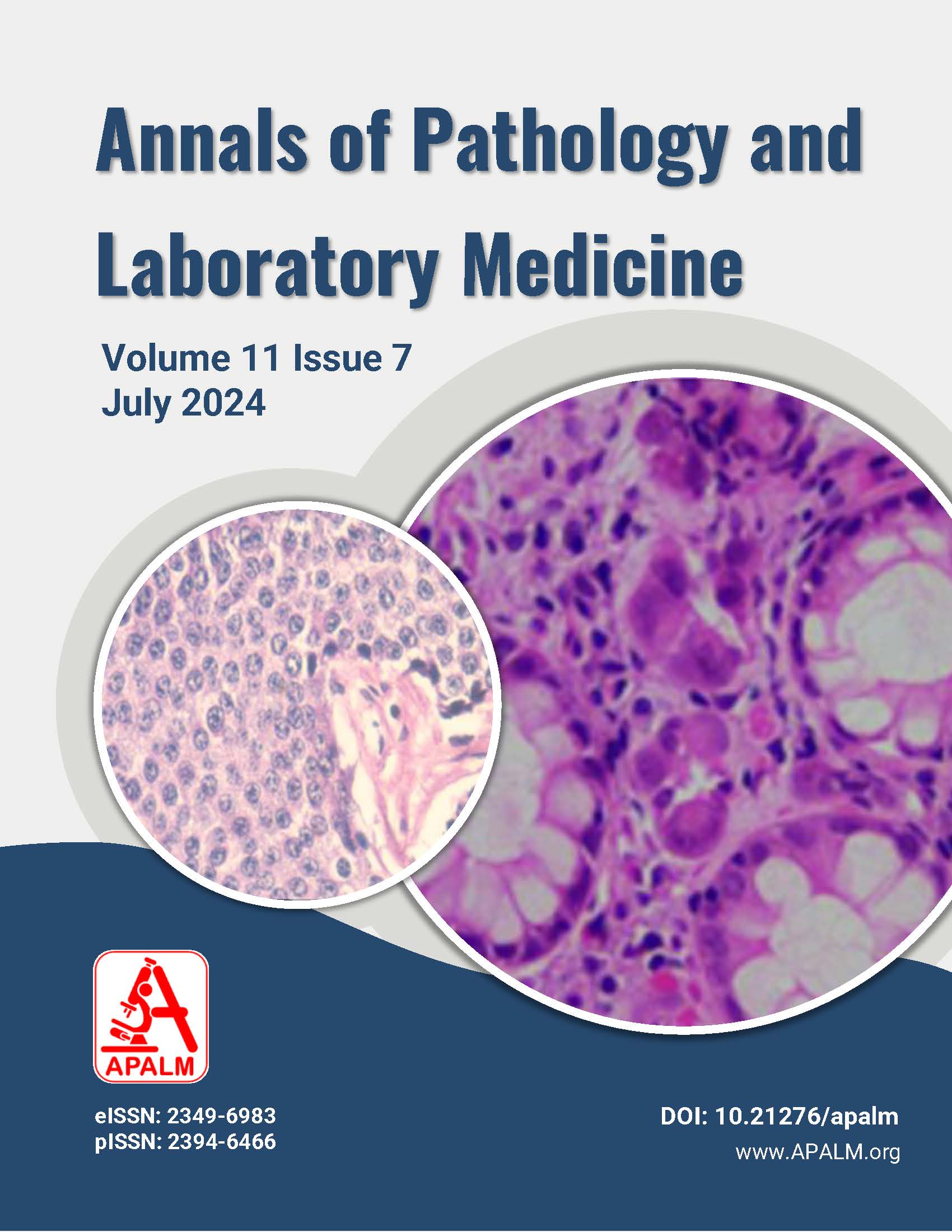
Case Report: We present a case of a 30-year-old female with symptoms of giddiness, focal convulsions, and loss of consciousness, along with positive cerebellar signs. On MRI, an ill-defined intra-axial lesion showed classical alternating light and dark "tiger stripe" patterns with no contrast enhancement. Frozen and histopathological evaluations confirmed the diagnosis, as there was the presence of atypical ganglion cells replacing the internal molecular layer. The patient underwent total tumor resection.
Conclusion: Lhermitte-Duclos disease is a rare lesion that unilaterally enlarges the cerebellum but maintains the foliar architecture. It is identified as a component of Cowden syndrome, an autosomal dominant cancer predisposition disorder. Recognition of the histological characteristics of this uncommon condition and maintaining a heightened suspicion are imperative for an accurate diagnosis. This should prompt comprehensive examinations to rule out manifestations of associated Cowden syndrome. Lhermitte-Duclos Disease is an infrequent occurrence, and understanding its histological attributes and aligning them with radiological findings is crucial, particularly in small biopsy samples, to ensure a precise diagnosis.
References
Cheng C-S, Aygun N, Lee LH, McCarthy EF, Westra WH. Lhermitte-Duclos disease: A case report with radiologic-pathologic correlation. Radiol Case Rep. 2019 Apr 3;14(6):734-9.
Kolhe AA, Shenoy A, Tayal S, Goel NA. Lhermitte-Duclos disease: a series of six cases. J Neurosci Rural Pract. 2023;14:127-31.
Nowak DA, Trost HA. Lhermitte-Duclos disease (dysplastic cerebellar gangliocytoma): A malformation, hamartoma or neoplasm? Acta Neurol Scand. 2002;105:137-45.
Zhou XP, Marsh DJ, Morrison CD, Chaudhury AR, Maxwell M, Reifenberger G, et al. Germline inactivation of PTEN and dysregulation of the phosphoinositide-3-kinase/Akt pathway cause human Lhermitte-Duclos disease in adults. Am J Hum Genet. 2003;73:1191-8.
Thomas B, Krishnamoorthy T, Radhakrishnan VV, Kesavadas C. Advanced MR imaging in Lhermitte-Duclos disease: Moving closer to pathology and pathophysiology. Neuroradiology. 2007;49:733-8.
Kulkantrakorn K, Awwad EE, Levy B, Selhorst JB, Cole HO, Leake D. MRI in Lhermitte-Duclos disease. Neurology. 1997;48:725-31.
Eberhart CG, Wiestler OD, Eng C. Dysplastic cerebellar gangliocytoma (Lhermitte-Duclos disease). In: Louis DN, editor. WHO Classification of Tumors of the Central Nervous System. 4th ed. Lyon: IARC; 2016. p. 142-3.
Pérez-Núñez A, Lagares A, BenÃtez J, Urioste M, Lobato RD, Ricoy JR, et al. Lhermitte-Duclos disease and Cowden disease: Clinical and genetic study in five patients with Lhermitte-Duclos disease and literature review. Acta Neurochir (Wien). 2004;146:679-90.
Downloads
Published
Issue
Section
License
Copyright (c) 2024 Charu Kiran Agrawal, Nitin M Gadgil, Chetan Chaudhari, Aishwarya Yedke

This work is licensed under a Creative Commons Attribution 4.0 International License.
Authors who publish with this journal agree to the following terms:
- Authors retain copyright and grant the journal right of first publication with the work simultaneously licensed under a Creative Commons Attribution License that allows others to share the work with an acknowledgement of the work's authorship and initial publication in this journal.
- Authors are able to enter into separate, additional contractual arrangements for the non-exclusive distribution of the journal's published version of the work (e.g., post it to an institutional repository or publish it in a book), with an acknowledgement of its initial publication in this journal.
- Authors are permitted and encouraged to post their work online (e.g., in institutional repositories or on their website) prior to and during the submission process, as it can lead to productive exchanges, as well as earlier and greater citation of published work (See The Effect of Open Access at http://opcit.eprints.org/oacitation-biblio.html).
How to Cite


