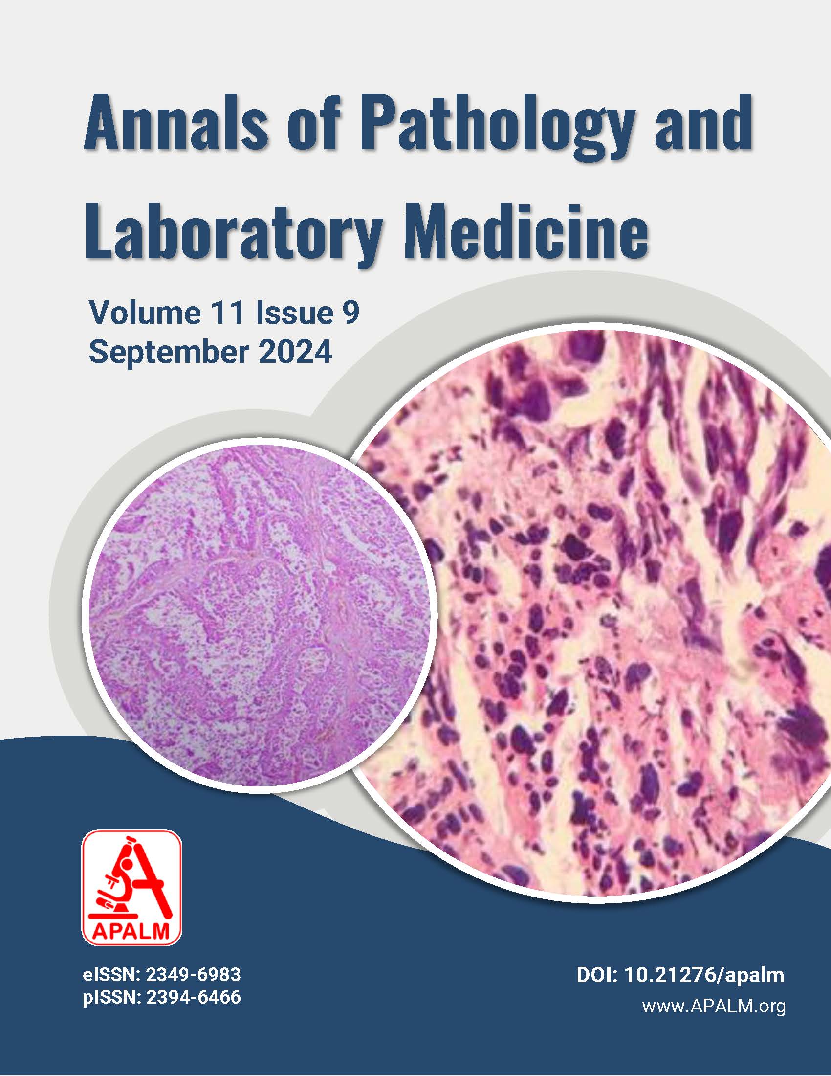
A Highly Aggressive Choriocarcinoma of Tubo-Ovarian Mass: A Rare Case Report
DOI:
https://doi.org/10.21276/apalm.3384Keywords:
Choriocarcinoma, Gestational, Nongestational, Tubo-Ovarian massAbstract
Background: Choriocarcinoma is a rare and highly aggressive tumor of trophoblastic origin. The most common location of choriocarcinoma is intrauterine; however, rare sites include the fallopian tube, ovary, and elsewhere in the abdomen and pelvis.
Case details: A 46-year-old woman, with a gestational history of G5P5Sb5, presented to the gynecology OPD with complaints of abdominal pain for four to six (4—6) months and a history of irregular menstrual periods. CECT chest and abdomen revealed a large left adnexal mass, moderate ascites, and pleural effusion. Serial beta-HCG levels were elevated. Specimens of the uterus, right salpingo-oophorectomy, and left adnexal mass were received for histopathological examination. On gross examination, there was a grayish, irregular left adnexal tissue mass measuring 20 × 14 × 9.0 cm. On cut section, multiple necrotic, hemorrhagic, and cystic areas were noted. No gross abnormalities were seen in the uterus, right fallopian tube, and right ovary. Multiple sections studied from the en masse tissue showed sheets of atypical syncytiotrophoblast and cytotrophoblast with two to three (2—3) mitotic figures per ten (10) high-power fields. The tumor showed an infiltrative and destructive pattern with an extensive necrotic and hemorrhagic background.
Conclusion: Choriocarcinoma is a rare but highly aggressive tumor. It usually presents with symptoms resulting from metastasis to the lungs, central nervous system, or alimentary tract. In our case, considering the significant clinical history, high levels of beta-HCG, and histopathological findings, it was diagnosed as a case of choriocarcinoma of the left tubo-ovarian mass.
References
Desai NR, Gupta AS, Mokal R, Jhaveri MB. Choriocarcinoma in a 73-year-old woman: a case report and review of literature. J Med Case Rep. 2010;4:20.
Bishop BN, Edemekong PF. Choriocarcinoma. Treasure Island (FL): StatPearls Publishing; 2024 Jan.
Corakci A, Ozeren S, Gurbuz Y, Ustun H, Yucesoy I. Pure nongestational choriocarcinoma of ovary. Arch Gynecol Obstet. 2005;271(2):176-9.
Koo HL, Choi J, Kim KR, Kim JH. Pure nongestational choriocarcinoma of the ovary diagnosed by DNA polymorphism. Pathol Int. 2006;56:34-9.
Yamamoto E, Ino K, Yamamoto T, Sumigama S, Nawa A, Nomura S, et al. A pure non-gestational choriocarcinoma of the ovary diagnosed with short tandem repeat analysis: case report and review of literature. Int J Gynecol Cancer. 2007;17(1):254-8.
Downloads
Published
Issue
Section
License
Copyright (c) 2024 Shahnaz Bano, Bhanu Pratap Singh, Nilopher Hirani

This work is licensed under a Creative Commons Attribution 4.0 International License.
Authors who publish with this journal agree to the following terms:
- Authors retain copyright and grant the journal right of first publication with the work simultaneously licensed under a Creative Commons Attribution License that allows others to share the work with an acknowledgement of the work's authorship and initial publication in this journal.
- Authors are able to enter into separate, additional contractual arrangements for the non-exclusive distribution of the journal's published version of the work (e.g., post it to an institutional repository or publish it in a book), with an acknowledgement of its initial publication in this journal.
- Authors are permitted and encouraged to post their work online (e.g., in institutional repositories or on their website) prior to and during the submission process, as it can lead to productive exchanges, as well as earlier and greater citation of published work (See The Effect of Open Access at http://opcit.eprints.org/oacitation-biblio.html).


