
Histopathological Spectrum of Splenectomy Specimens: A Six-Year Retrospective Study
DOI:
https://doi.org/10.21276/apalm.3563Keywords:
Splenectomy, chronic venous congestion, traumatic lacerationAbstract
Background: The spleen plays a vital role in blood filtration and immune function but can be affected by a range of conditions requiring surgical removal. This study explores the histopathological findings in splenectomy specimens received at our institute and examines how these findings relate to clinical diagnosis.
Methods: We conducted a retrospective study over six years (2016-2021) at the Department of Pathology, SDM College of Medical Sciences and Hospital, Karnataka. A total of 60 splenectomy specimens were analyzed through detailed gross and microscopic examination, with clinical data retrieved from medical records.
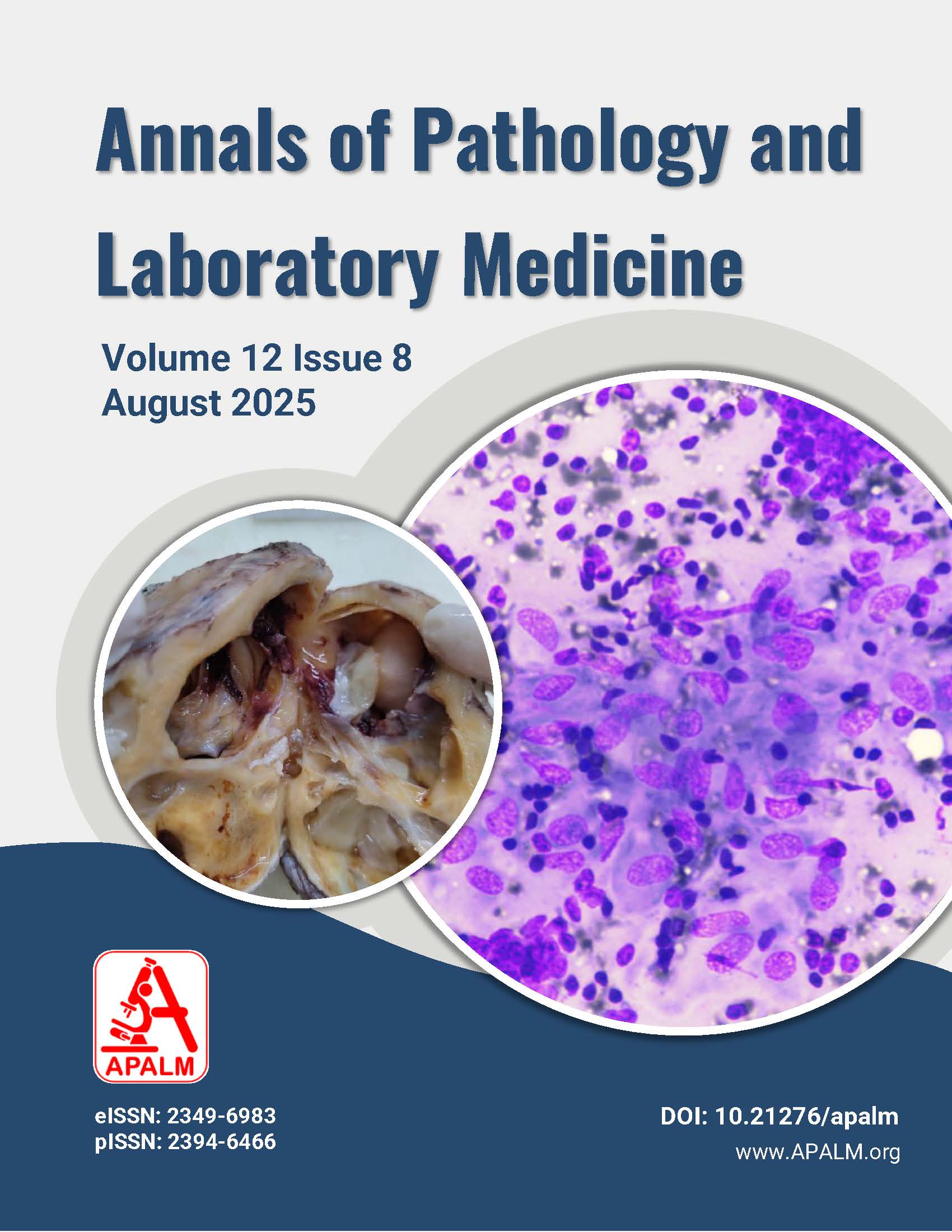
Results: Of the 60 patients, 36 were male and 24 were female, with the most common age group being 31-40 years. The leading reason for splenectomy was blunt abdominal trauma (40%), followed by blood disorders like thalassemia intermedia (11.7%) and hypersplenism (8.3%). Histopathological examination most frequently revealed traumatic laceration (38.3%) and chronic venous congestion (36.7%), often linked to conditions like thalassemia and portal hypertension. Less common findings included splenic hamartomas, lymphangiomas, and Non-Hodgkin's lymphoma. Cystic lesions were identified in 10% of cases, including pseudocysts, mesothelial cysts, and epithelial cysts.
Conclusion: Although splenectomy is not a routine procedure, its histopathological analysis provides valuable insights into underlying diseases. This study highlights trauma as the most common reason for spleen removal, followed by hematological and vascular disorders. Understanding these patterns can help improve diagnosis and patient management.
References
1. Chary R, Fatima A, Husain KW, Chalam PK, Mohammed I. Spectrum of splenic pathology at a single centre. Indian Journal of Pathology and Oncology. 2016 Jan 1;3(4):622.
2. Chaudhry SR, Luskin V, Panuganti KK. Anatomy. Abdomen and Pelvis, Spleen. In: StatPearls. Treasure Island (FL): StatPearls Publishing; July 24, 2023.
3. Mahadevan V. Anatomy of the pancreas and spleen. Surgery (Oxford). 2019 Jun 1:37(6):297-301.
4. Cha A. The spleen: Anatomy and anatomical function. Semin Hematol. 2000;37(Suppl 1):13-21.
5. Cesta MF. Normal structure, function, and histology of the spleen. Toxicol Pathol. 2006:34:455-465.
6. Goldblum JR, Lamps LW, McKenney JK, Myers JL. Rosai and Ackerman's Surgical Pathology. 11th ed. Philadelphia Elsevier 2018.
7. Nigudgi S, Sheelwanth S. Anita AM, Patil A, Andola SK. Spectrum of rare splenic lesions. J Diagn Pathol Oncol. 2018;3(3):223-227.
8. Banerjee A, Datta A, Das S. Histomorphological spectrum of splenectomy specimens in a tertiary teaching hospital: A seven year study. Int J Med and Dent Sci 2017:6(2):1488-1492.
9. Choudhary M, Jandial R. Histopathological review of splenectomy specimens: A five-year study in a tertiary care centre in North India. Int J Health Sci Res. 2019;9(10):242-248.
10. Adelusola KA, Osasan SA, Afolabi OA. Histopathological study and audit of the spleen in Nigerians. Afr J Health Sci. 2007;14(3-4):195-202.
11. Namratha R, Vijaya B, Bhadran K. A histopathological study of spectrum of splenic lesions: An eleven-year analysis of clinical and pathological aspects of splenectomy specimens in a tertiary care hospital. Indian J Pathol Oncol. 2022 9(3):220-226.
12. Gupta D, Gupta A, Suri J, Singh K. Histomorphological spectrum in splenectomy specimens: A five-year retrospective study. Indian J Pathol Oncol. 2017 4(3):418-421.
13. Sarangthem B. Pukhrambam GD, Laishram S, Sharma AB, Debnath K. Histomorphological pattern of splenectomy specimens: A five-year study in a tertiary teaching hospital. IOSR J Dent Med Sci. 2014;13(1):40-43.
14. Jagadev S, Balaji C, Chappa S, et al. Histomorphological pattern of splenectomy specimens a two year study in a tertiary teaching hospital. J Evid Based Med Healthc 2020; 7(35),1867-1870.
Downloads
Published
Issue
Section
License
Copyright (c) 2025 Basavaraj Dundappa Yamakanamardi, Deepti Shivananad Shettar, Malashree, Aneel Myageri

This work is licensed under a Creative Commons Attribution 4.0 International License.
Authors who publish with this journal agree to the following terms:
- Authors retain copyright and grant the journal right of first publication with the work simultaneously licensed under a Creative Commons Attribution License that allows others to share the work with an acknowledgement of the work's authorship and initial publication in this journal.
- Authors are able to enter into separate, additional contractual arrangements for the non-exclusive distribution of the journal's published version of the work (e.g., post it to an institutional repository or publish it in a book), with an acknowledgement of its initial publication in this journal.
- Authors are permitted and encouraged to post their work online (e.g., in institutional repositories or on their website) prior to and during the submission process, as it can lead to productive exchanges, as well as earlier and greater citation of published work (See The Effect of Open Access at http://opcit.eprints.org/oacitation-biblio.html).
How to Cite


