
Immunohistochemical Expression of CK19 and CD56 in Differentiating Malignant Neoplasms of Thyroid from Its Benign Mimickers
DOI:
https://doi.org/10.21276/apalm.3570Keywords:
Immunohistochemistry, CK19, CD56, thyroid neoplasmsAbstract
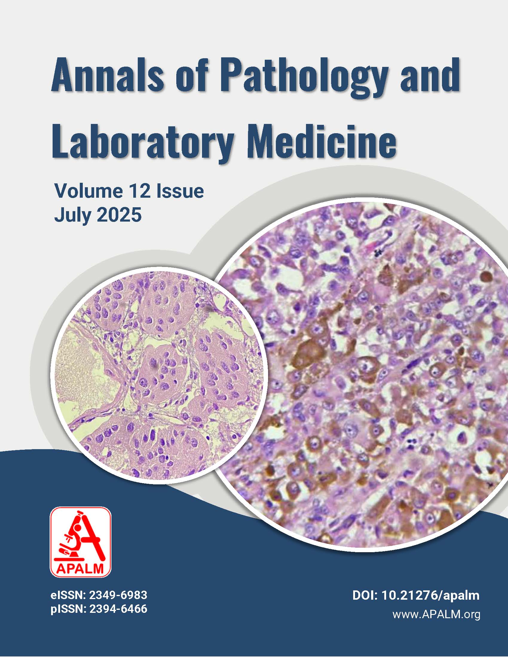
Background: Papillary formation is often observed in benign and malignant thyroid diseases, meaning that it is difficult to distinguish between benign and malignant lesions; hence, diagnosis based only on histopathology is often challenging, even in the hands of experienced pathologists. Thus, immunohistochemistry (IHC) is also essential in the diagnosis. This study aims to observe the utility of IHC markers (CK19 and CD56) in thyroid neoplasms.
Methods: This cross-sectional study was conducted in the Department of Pathology at Bhagat Phool Singh Government Medical College for Women, Khnapur Kalan, Sonipat, Haryana. A total of 55 specimens, including 27 benign neoplasms and 28 malignant neoplasms, were subjected to IHC (CD56 and CK19).
Results: Out of 55 thyroid neoplasm cases, 27 were benign and 28 were malignant. Immunohistochemistry CK19 was positive in 78.5% (22/28) of malignant cases, while negative in 78% (21/27) of benign cases with a significant p-value of <0.001; while immunohistochemistry for CD56 was positive in 92.6% (25/27) of benign cases, and 67.9% (19/28) of malignant cases showed loss of CD56 expression with a highly significant p-value of <0.001. The expression of CK19 was found to have 75% sensitivity and 74.1% specificity, while loss of CD56 expression was found to have 90.4% sensitivity and 73.5% specificity in differentiating malignant from benign thyroid neoplasms.
Conclusion: CK19 is a specific marker for thyroid cancer, especially useful for identifying PTC and FVPTC. CD56 serves as a sensitive marker, with loss of expression indicating malignant potential, enhancing diagnostic accuracy in ambiguous cases.
References
1. Maitra A. The endocrine system. In: Kumar V, Abbas AK, Aster JC, Turner JR, Singh MK, editors. Robbins and Cotran pathologic basis of disease, 10th ed. Philadelphia: Elsevier; 2018. р. 1075-92.
2. Thyroid Globocan-2022: World-factsheet. Available from: https://gco.iarc.fr/today/data/factsheets/cancers/32-Thyroid-fact-sheet.pdf.
3. Huang L, Wang X, Huang X, Gui H, Li Y, Chen Q, et al. Diagnostic significance of CK19, galectin-3, CD56, TPO and Ki67 expression and BRAF mutation in papillary thyroid carcinoma. Oncol Lett. 2018;15:4269-77.
4. Dwivedi SS, Khandeparkar SG, Joshi AR, Kulkarni MM, Bhayekar P, Jadhav A, et al. Study of immunohistochemical markers (CK-19, CD-56, Ki-67, p53) in differentiating benign and malignant solitary thyroid nodules with special reference to papillary thyroid carcinomas. J Clin Diagn Res. 2016;10(12):14-9.
5. Tastekin E, Keskin E, Can N, Canberk S. Mut A, Erdogan E, et al. CD56, CD57, HBME1, CK19, Galectin-3 and p63 immunohistochemical stains in differentiating diagnosis of thyroid benign/malign lesions and NIFTP. Pol J Pathol. 2019:70(4):286-94.
6. Kumari AS, Sinha P. Role of CD56 and cytokeratin 19 in the diagnosis of papillary and follicular neoplasms of thyroid. High Technol Lett. 2020 29(9):842-50.
7. Zughaibi IF, Kamal MS. The part immunohistochemical markers (CK19 and CD56) play in distinguishing papillary thyroid carcinoma from other pathological imitators. Egypt J Hosp Med. 2023;90(2):3495-500.
8. Raman J. Philip FK, Govind U. Diagnostic utility of immunohistochemistry markers galectin-3, CK19 and CD56 in thyroid neoplasms: a descriptive study. J Clin Diagn Res. 2022;16(12):28-33.
9. Dalal N, Sharma U, Sweta, Baki IK. Application of CK19 and CD56 immunohistochemical markers in the diagnosis of thyroid tumors. J med sci clin res. 2018;6(2):1118-27.
10. Kammal WS, Yahaya A, Shah SA, Suhaimi SN, Mahasin M, Mustangin M, et al. The diagnostic utility of cytokeratin 19 in differentiating malignant from benign thyroid lesions. Malays J Pathol. 2019;41(3):293-301.
11. Priyadarshini P, Patro MK, Das PK. Diagnostic role of immunohistochemical markers CK19 and CD56 in thyroid neoplasms. MGM J Med Sci. 2023;10(2):176-83.
12. Dunerović D. Lipkovski JM, Boričic I, Soldatović I, Božic V, Cvejić D, et al. Defining the value of CD56, CK19, Galectin 3 and HBME-1 in diagnosis of follicular cell derived lesions of thyroid with systematic review of literature. Diagn Pathol. 2015;10:1-18.
13. Muthusamy S, Sha SA, Suhaimi SN, Kassim N, Mahasin M, Saleh MF, et al. CD56 expression in benign and malignant thyroid lesions. Malays J Pathol. 2018 Aug:40(2):111-9.
Downloads
Published
Issue
Section
License
Copyright (c) 2025 Tanu Priya, Monika B Gathwal, Swaran Kaur, Vijyendra Puri Goswami, Chiranjeev Gathwal

This work is licensed under a Creative Commons Attribution 4.0 International License.
Authors who publish with this journal agree to the following terms:
- Authors retain copyright and grant the journal right of first publication with the work simultaneously licensed under a Creative Commons Attribution License that allows others to share the work with an acknowledgement of the work's authorship and initial publication in this journal.
- Authors are able to enter into separate, additional contractual arrangements for the non-exclusive distribution of the journal's published version of the work (e.g., post it to an institutional repository or publish it in a book), with an acknowledgement of its initial publication in this journal.
- Authors are permitted and encouraged to post their work online (e.g., in institutional repositories or on their website) prior to and during the submission process, as it can lead to productive exchanges, as well as earlier and greater citation of published work (See The Effect of Open Access at http://opcit.eprints.org/oacitation-biblio.html).
How to Cite


