
Fine-Needle Aspiration Cytology of Extramedullary Plasmacytoma: A Six-Year Case Series
DOI:
https://doi.org/10.21276/apalm.3622Keywords:
Extramedullary plasmacytoma, Fine needle aspiration cytology, Plasmablast, Light chain restriction, RadiotherapyAbstract
Background: Due to its cytomorphological overlap with lymphoma or metastatic carcinoma and its protean anatomic distribution, extramedullary plasmacytoma (EMP), which accounts for less than 5% of plasma cell neoplasms, usually presents a diagnostic problem. Timely staging and treatment depend on early cytological identification.
Materials and Methods: We retrospectively reviewed 8 cases of extramedullary plasmacytoma (EMP) diagnosed on fine needle aspiration cytology (FNAC). Clinical history, imaging, laboratory data, treatment, and follow-up were retrieved from hospital records. FNAC smear samples were assessed. Cell blocks were prepared in all 8 cases and subjected to immunocytochemistry for CD138, CD38, CD45, and Ki-67 markers. In situ hybridization for κ and λ light chains was done in all cases. Descriptive analysis was performed given the small sample size.
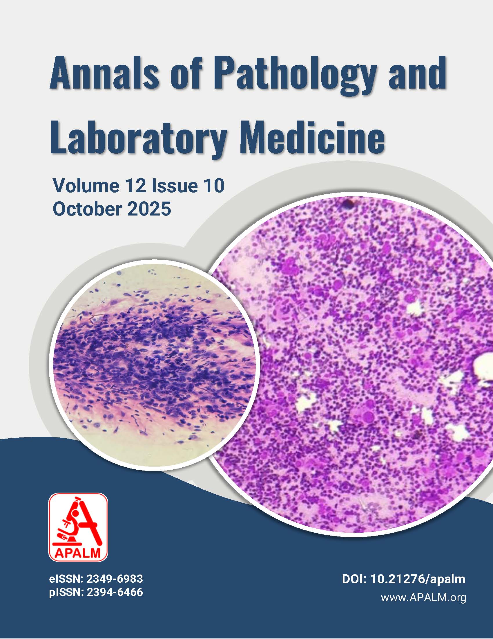
Results: Eight patients (mean age 70.3 years; M:F=5:3) presented with solitary soft tissue masses ranging from 2.8 to 6.5 cm. Sites included paranasal sinus (2), axilla (1), breast (1), sternum (1), orbit (1), scapula (1), and subcutaneous thigh (1). FNAC smears showed a spectrum from mature plasma cells to plasmablast-rich populations. Immunocytochemistry revealed monotypic κ-restriction in five cases and λ in three. Over a median follow-up of 15.5 months, five patients remained disease-free, two progressed to multiple myeloma, and one was lost to follow-up.
Conclusion: FNAC, combined with light-chain assessment and meticulous clinico-radiological correlation, is a rapid and reliable modality for diagnosing EMP even in resource-constrained settings. Recognizing plasmablast-rich patterns is essential because they herald aggressive behavior and a higher risk of progression to myeloma.
References
1. Alexiou C, Kau RJ, Dietzfelbinger H, Kremer M, Spieß JC, Schratzenstaller B, et al. Extramedullary plasmacytoma: tumor occurrence and therapeutic concepts. Cancer. 1999;85(11):2305-14.
2. Lai CY, Hsieh HH, Chen HK, Chao CY, Hua CH, et al. Clinical Features of Head and Neck Solitary Extramedullary Plasmacytoma in Taiwan. In Vivo, 2020;34(1):261-5. doi: 10.21873/invivo. 11769.
3. Harmon CM, Smith LB. Plasmablastic Lymphoma: A Review of Clinicopathologic Features and Differential Diagnosis. Arch Pathol Lab Med. 2016;140(10):1074-8. doi: 10.5858/arpa.2016-0232-RA.
4. Castillo JJ, Bibas M, Miranda RN. The biology and treatment of plasmablastic lymphoma. Blood. 2015;125(15):2323-30. doi: 10.1182/blood-2014-10-567479.
5. Greipp PR, San Miguel J, Durie BG, Crowley JJ, Barlogie B, Bladé J, et al. International staging system for multiple myeloma. J Clin Oncol. 2005;23(15):3412-20. doi: 10.1200/JCO.2005.04.242.
6. Dimopoulos MA, Moreau P, Terpos E, Mateos MV, Zweegman S, Cook G, et al. Multiple myeloma: EHA-ESMO Clinical Practice Guidelines for diagnosis, treatment and follow-up. Hemasphere. 2021;32(3):309-22. doi: 10.1016/j.annonc.2021.10.001.
7. Tsang RW, Campbell BA, Goda JS, Kelsey CR, Kirova YM, et al. Radiation Therapy for Solitary Plasmacytoma and Multiple Myeloma: Guidelines From the International Lymphoma Radiation Oncology Group. Int J Radiat Oncol Biol Phys. 2018:101(4):794-808. doi: 10.1016/j.ijrobp.2018.05.009.
8. Bartl R, Frisch B, Burkhardt R. Fateh-Moghadam A, Mahl G, et al. Bone marrow histology in myeloma: its importance in diagnosis, prognosis, classification and staging. Br J Haematol. 1982;51(3):361-75. doi: 10.1111/j.1365-2141.1982.tb02791.x.
9. Sabattini E, Bacci F, Sagramoso C, Pileri SA. WHO classification of tumours of haematopoietic and lymphoid tissues in 2008: an overview. Pathologica. 2010;102(3):83-7.
10. Rawstron AC, Child JA, de Tute RM, Davies FE, Gregory WM, et al. Minimal residual disease assessed by multiparameter flow cytometry in multiple myeloma: impact on outcome in the Medical Research Council Myeloma IX Study. J Clin Oncol. 2013 31(20):2540-7. doi: 10.1200/JCO.2012.46.2119.
11. Goel G, Rai S, Naik R, Gupta A, Baliga P, Sinha R. Cytodiagnosis of extramedullary plasmacytomas. Acta Cytol. 2010;54(3):255-8. doi: 10.1159/000325031.
12. Arber DA, Orazi A, Hasserjian R, Thiele J, Borowitz MJ, et al. The 2016 revision to the World Health Organization classification of myeloid neoplasms and acute leukemia. Blood. 2016;127(20):2391-405.
13. Dimopoulos MA, Kiamouris C, Moulopoulos LA. Solitary plasmacytoma of bone and extramedullary plasmacytoma. Hematol Oncol Clin North Am. 1999;13(6):1249-57. doi: 10.1016/s0889-8588(05)70124-6.
14. Grammatico S, Scalzulli E, Petrucci MT. Solitary plasmacytoma. Mediterranean journal of hematology and infectious diseases. 2017:9(1):e2017052.
15. Sher T, Miller KC, Deeb G, Lee K, Chanan-Khan A. Plasma cell leukaemia and other aggressive plasma cell malignancies. British journal of haematology. 2010;150(4):418-27.
Downloads
Published
Issue
Section
License
Copyright (c) 2025 Tripti Jain, Surinder Singh, Arsha Narayanan, Sujata Kumari, Harinder Singh Chhabra

This work is licensed under a Creative Commons Attribution 4.0 International License.
Authors who publish with this journal agree to the following terms:
- Authors retain copyright and grant the journal right of first publication with the work simultaneously licensed under a Creative Commons Attribution License that allows others to share the work with an acknowledgement of the work's authorship and initial publication in this journal.
- Authors are able to enter into separate, additional contractual arrangements for the non-exclusive distribution of the journal's published version of the work (e.g., post it to an institutional repository or publish it in a book), with an acknowledgement of its initial publication in this journal.
- Authors are permitted and encouraged to post their work online (e.g., in institutional repositories or on their website) prior to and during the submission process, as it can lead to productive exchanges, as well as earlier and greater citation of published work (See The Effect of Open Access at http://opcit.eprints.org/oacitation-biblio.html).
How to Cite


