
Histomorphological Changes and Pathological Response Following Neoadjuvant Chemotherapy in Breast Carcinoma: A Cross-Sectional Study
DOI:
https://doi.org/10.21276/apalm.3818Keywords:
breast carcinoma, neoadjuvant chemotherapy, histomorphology, Miller–Payne grading, pathological responseAbstract
Background: Neoadjuvant chemotherapy (NACT) is widely used in the management of locally advanced breast carcinoma to downstage tumors and improve surgical outcomes. Histopathological evaluation of post-NACT specimens and assessing pathological response using the Miller–Payne grading system provides critical information regarding therapeutic response and prognosis.
Methods: This cross-sectional study included 50 patients with biopsy-proven breast carcinoma who received two or more cycles of neoadjuvant chemotherapy followed by surgical excision. Post-therapy specimens were examined for nuclear, stromal, and inflammatory changes using routine hematoxylin and eosin staining. Tumor regression was graded according to the Miller–Payne system. Statistical analysis was performed using SPSS version 20, with p < 0.05 considered statistically significant.
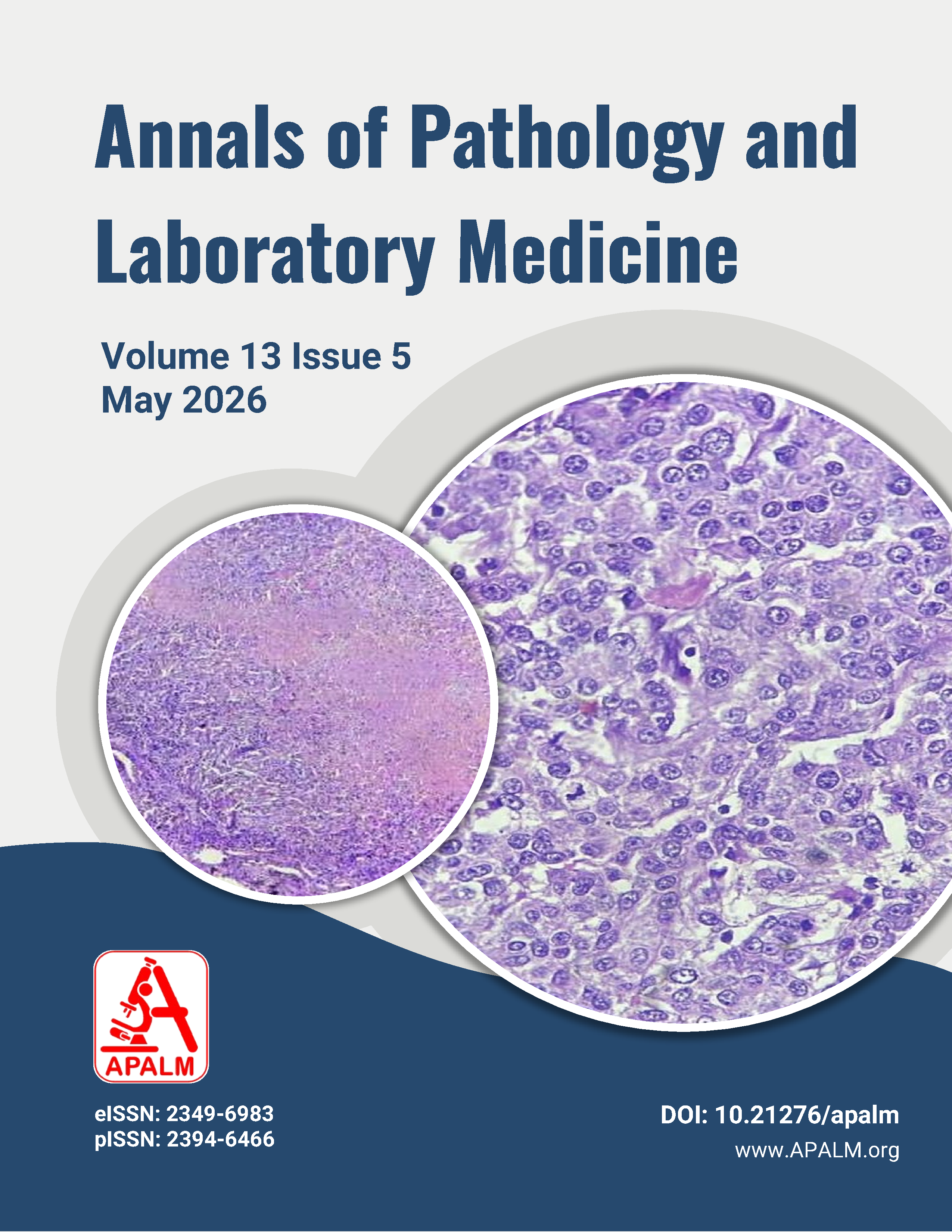
Result: The mean age of patients was 52.78 ± 10.43 years. Invasive ductal carcinoma, no special type, was the most common histological subtype (88%). A significant reduction in tumor size was observed following neoadjuvant chemotherapy, with mean tumor size decreasing from 4.87 ± 1.14 cm pre-NACT to 1.94 ± 1.16 cm post-NACT (p < 0.0001). Histomorphological changes included nuclear alterations in 60% of cases, stromal fibrosis or sclerosis in 52%, tumor necrosis in 64%, and inflammatory infiltrate in 90%. Pathological complete response was achieved in 14% of cases, while partial response was observed in 64%.
Conclusion: Histopathological evaluation remains the most reliable method for assessing response to neoadjuvant chemotherapy in breast carcinoma. Therapy-induced nuclear and stromal changes correlate with pathological response and have important prognostic implications.
References
1. Bray F, Ferlay J, Soerjomataram I, Siegel RL, Torre LA, Jemal A. Global cancer statistics 2018: GLOBOCAN estimates of incidence and mortality worldwide. CA Cancer J Clin. 2018;68(6):394–424.
2. Early Breast Cancer Trialists' Collaborative Group (EBCTCG). Effects of chemotherapy and hormonal therapy for early breast cancer on recurrence and survival. Lancet. 2005;365(9472):1687–1717.
3. Fisher B, Brown A, Mamounas E, et al. Effect of preoperative chemotherapy on local-regional disease in women with operable breast cancer. J Clin Oncol. 1997;15(7):2483–2493.
4. Provenzano E, Bossuyt V, Viale G, et al. Standardization of pathologic evaluation of residual disease after neoadjuvant therapy. Mod Pathol. 2015;28(9):1185–1201.
5. Bi C, Chen A, Ran F, Hu Z, Sun S, Wang R, Niu X, Deng L, Gao D, Li Q, Yang J. Pretreatment MRI radiomics for predicting pathological Miller-Payne grading in breast cancer following neoadjuvant chemotherapy. Cancer Imaging. 2026 Jan 16.
6. Symmans WF, Peintinger F, Hatzis C, et al. Measurement of residual breast cancer burden to predict survival after neoadjuvant chemotherapy. J Clin Oncol. 2007;25(28):4414–4422.
7. von Minckwitz G, Untch M, Blohmer JU, et al. Definition and impact of pathological complete response on prognosis. J Clin Oncol. 2012;30(15):1796–1804.
8. Sahoo S, Lester SC. Pathology of breast carcinomas after neoadjuvant chemotherapy. Semin Diagn Pathol. 2009;26(4):273–285.
9. Gupta D, Sharma A, Suri V, et al. Histopathological changes following neoadjuvant chemotherapy in breast carcinoma. Indian J Pathol Microbiol. 2016;59(4):466–471.
10. Ogston KN, Miller ID, Payne S, et al. A new histological grading system to assess response to primary chemotherapy in breast cancer. Breast. 2003;12(5):320–327.
11. Tiwari K, Verma N. Post-chemotherapy changes in breast with evaluation of residual carcinoma burden. Asian Journal of Medical Sciences. 2024 May 1;15(5):161-7.
12. Ahuja S, G K, Zaheer S. Evaluation of histomorphological changes in breast cancer post-neoadjuvant chemotherapy. Indian Journal of Surgical Oncology. 2024 Jun;15(2):236-40.
Downloads
Published
Issue
Section
License
Copyright (c) 2026 Agrima Kamra, Jyotsna Bhateja, Pushpinder Kaur

This work is licensed under a Creative Commons Attribution 4.0 International License.
Authors who publish with this journal agree to the following terms:
- Authors retain copyright and grant the journal right of first publication with the work simultaneously licensed under a Creative Commons Attribution License that allows others to share the work with an acknowledgement of the work's authorship and initial publication in this journal.
- Authors are able to enter into separate, additional contractual arrangements for the non-exclusive distribution of the journal's published version of the work (e.g., post it to an institutional repository or publish it in a book), with an acknowledgement of its initial publication in this journal.
- Authors are permitted and encouraged to post their work online (e.g., in institutional repositories or on their website) prior to and during the submission process, as it can lead to productive exchanges, as well as earlier and greater citation of published work (See The Effect of Open Access at http://opcit.eprints.org/oacitation-biblio.html).
How to Cite


